
Lighting Up Cancer Detection: Exploring New Materials for Medical Scanners
Medical scans like PET are vital tools for detecting and diagnosing cancer. At the heart of these scanners are materials called scintillators, which light up when hit by radiation. In his PhD research at Aarhus University, Simon Peter Slot Jessen studied how new nanomaterials could be used to build better, faster scintillators, using laser light to mimic real-world ionizing radiation.
When doctors need to find cancer early or monitor how it responds to treatment, they often use PET scans or other advanced imaging methods. These tools rely on scintillators, special materials that emit light when exposed to ionizing radiation. When a patient is given a small dose of radioactive tracer, the scanner detects the radiation using these materials. The scintillator absorbs the radiation and instantly emits light, which is then converted into a digital image that shows what is happening inside the body.
To improve how clearly and quickly doctors can see these images, researchers are constantly looking for better scintillators. Materials that emit more light, respond faster, and can be made safely and cost-effectively. A promising direction for next-generation scintillators is the use of highly luminous semiconductor nanoparticles. However, testing these tiny particles with ionizing radiation is difficult because the high-energy radiation penetrates deep into materials, making it hard to study how individual nanoparticles respond. This makes it challenging to isolate and understand their behavior using traditional radiation-based methods. Simon’s work studies the behavior of these scintillators using laser pulses instead.
Key findings
- Laser-based testing is used to safely simulate radiation effects in lab settings
- Laser excitation enables the study of nanosized structures independently, unlike ionizing radiation which mainly probes the bulk material
- CdSe/CdS nanoplatelets show promising light yield for PET imaging
- Dense packing in thin films can reduce light output due to nonlinear optical effects
Testing Scintillators Without Ionizing Radiation
Instead of testing materials with ionizing radiation, Simon used short and intense laser pulses to simulate the same conditions. These ultrafast flashes of light are intense enough to create excitation densities similar to ionizing radiation, allowing researchers to study how much light a material can produce, known as light yield, without using radioactive sources.
The method uses:
- Light with enough energy to excite the material
- Very fast pulses (less than a trillionth of a second) to match the speed of real energy deposition
- High intensity to reach the same level of excitation density a material would experience in a medical scanner
This setup made it possible to evaluate how different materials might perform in a real imaging device.
Focus on Nanomaterials
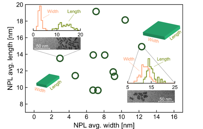
Simon concentrated on semiconductor nanomaterials, which are tiny crystals just a few billionths of a meter in size. One of the most promising types he studied was CdSe/CdS nanoplatelets. These have shown great potential for use in compact scintillator layers due to their strong and fast light emission. The dimensions of the nanometer sized plates that Simon studied are shown in Figure 1.
Using his laser-based setup, Simon studied thin films made from these nanoplatelets. He measured how much light they emitted when exposed to strong optical excitation and how their performance changed based on how they were arranged in the film.
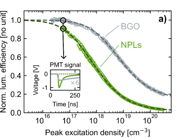
A key result was that CdSe/CdS nanoplatelets showed a light yield of around 2000 photons per MeV when tested under conditions that mimic real ionizing radiation exposure. This is a promising value compared to previous estimates using exctiation by ionizing radiation. The efficiency of the nanoplatelets (NPLs, green curve) approaches that of the commonly used scintillator material BGO (grey curve) in Figure 2.
However, the results Simon obtained also revealed an important challenge. When these nanoparticles are packed tightly into a film, just as they would be in an actual device, their performance can drop. This happens because the light-emitting particles can interfere with each other when they are too close, a process called nonlinear quenching. The result is less light output, even if the material is promising in theory.
This is a crucial insight for real-world applications, where scintillator materials are often packed into compact layers. This means that not only the type of material, but also how it is arranged, plays a big role in performance.
How Did They Do It?
To study the materials, Simon used:
- Ultrafast laser pulses to simulate ionizing radiation conditions
- Z-scan luminescence to measure how much light the samples emit
- Photoluminescence to analyze the quality and timing of the light output
- Film-based samples to reflect how materials would be used in real detectors
Why Does It Matter?
By studying how new nanomaterials behave under realistic energy conditions, Simon’s research helps researchers and engineers identify the best materials for future medical scanners. These could offer faster, more detailed images with lower doses of ionizing radiation, making cancer detection safer and more effective for patients.
In the long term, this work supports the development of more compact and affordable scanners.
Interested?
If you are working on related materials or applications, we invite you to reach out to our Center Manager to discuss potential collaborations or shared research opportunities.
You can read more here:
[1] Nonlinear quenching of excitonic emission from nanoplatelet films at high excitation densities
Simon Jessen, Alessio Di Giacomo, Iwan Moreels, Brian Julsgaard & Rosana M. Turtos
[1] Nonlinear quenching of excitonic emission from nanoplatelet films at high excitation densities
Simon Jessen, Alessio Di Giacomo, Iwan Moreels, Brian Julsgaard & Rosana M. Turtos